Cardiac Anatomy and Physiology
The heart is a major component of the cardiovascular system and plays a vital role in pumping blood throughout the body.
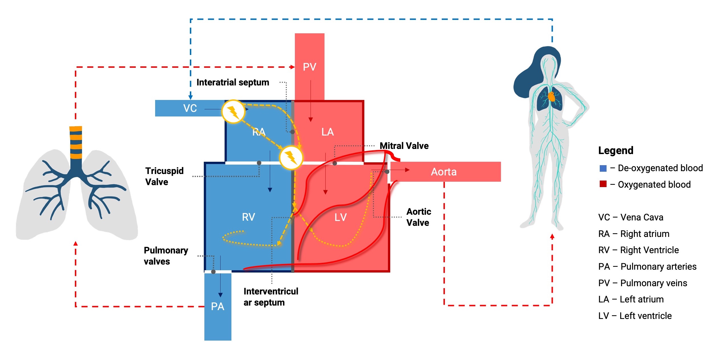
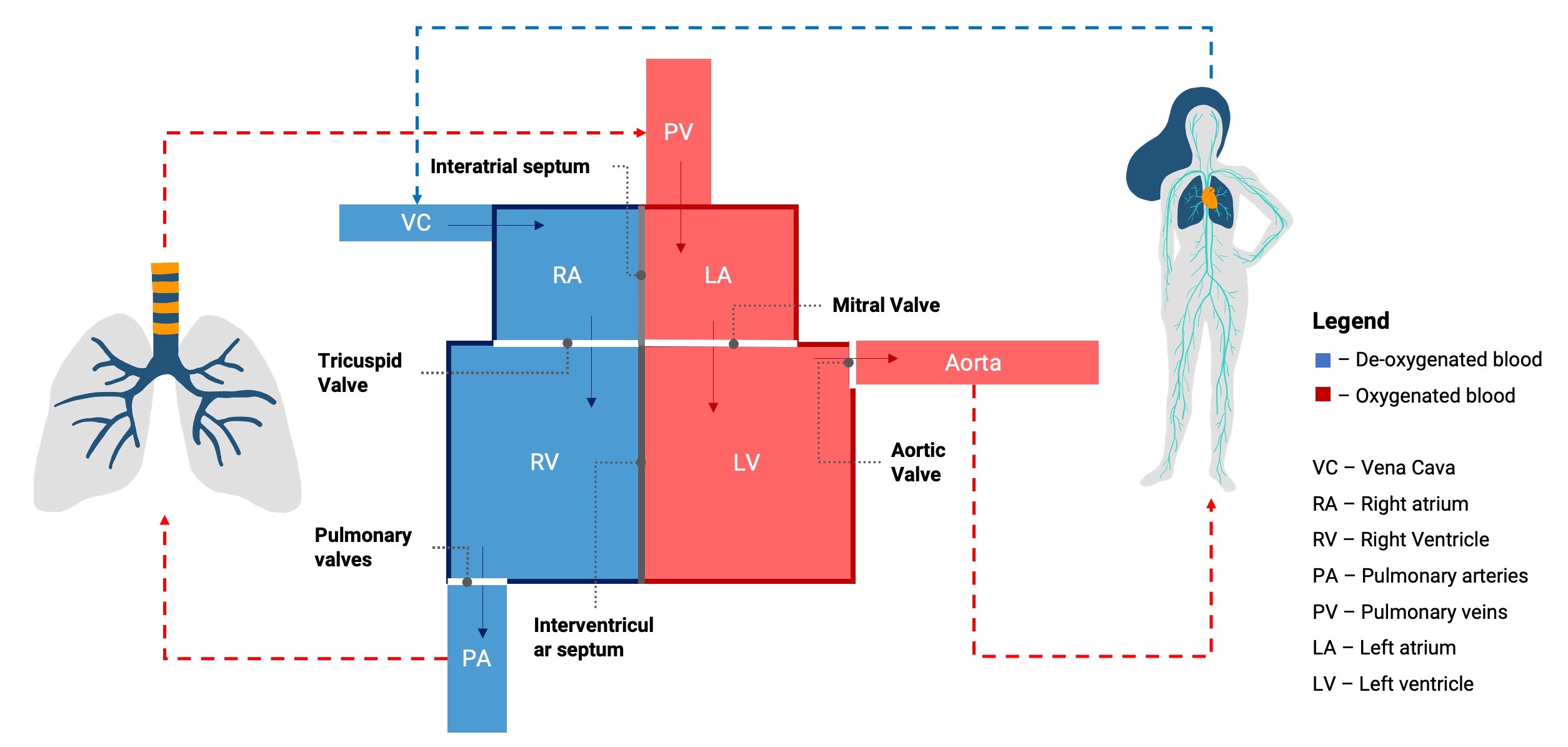
The heart contains:
- Septa
- Inter-atria septum
- Interventricular septum
- Layers of tissues
- Pericardium which is a thin outer lining which surrounds the heart
- Myocardium which is the muscular layer containing cardiac myocytes
- Endocardium which lines the inner chambers of the heart
- Chambers and Valves
- Right atrium which receives de-oxygenated blood from the body through the vena cava and pumps it across the tricuspid valve into the
- Right ventricle which pumps the blood into the lungs for oxygenation across the pulmonary valve via the pulmonary artery
- Left atrium which receives oxygenated blood from lungs through the pulmonary veins and moves it across the mitral valve into the
- Left ventricle which delivers blood to the rest of the body across the aortic valve via the aorta
The left ventricle is divided into different segments, determined by the distribution of the coronary artery. This is the septum, anterior, lateral, posterior and inferior walls (going clockwise from the septum). The region is also denoted by different electrocardiogram (ECG) leads on a 12-lead ECG.
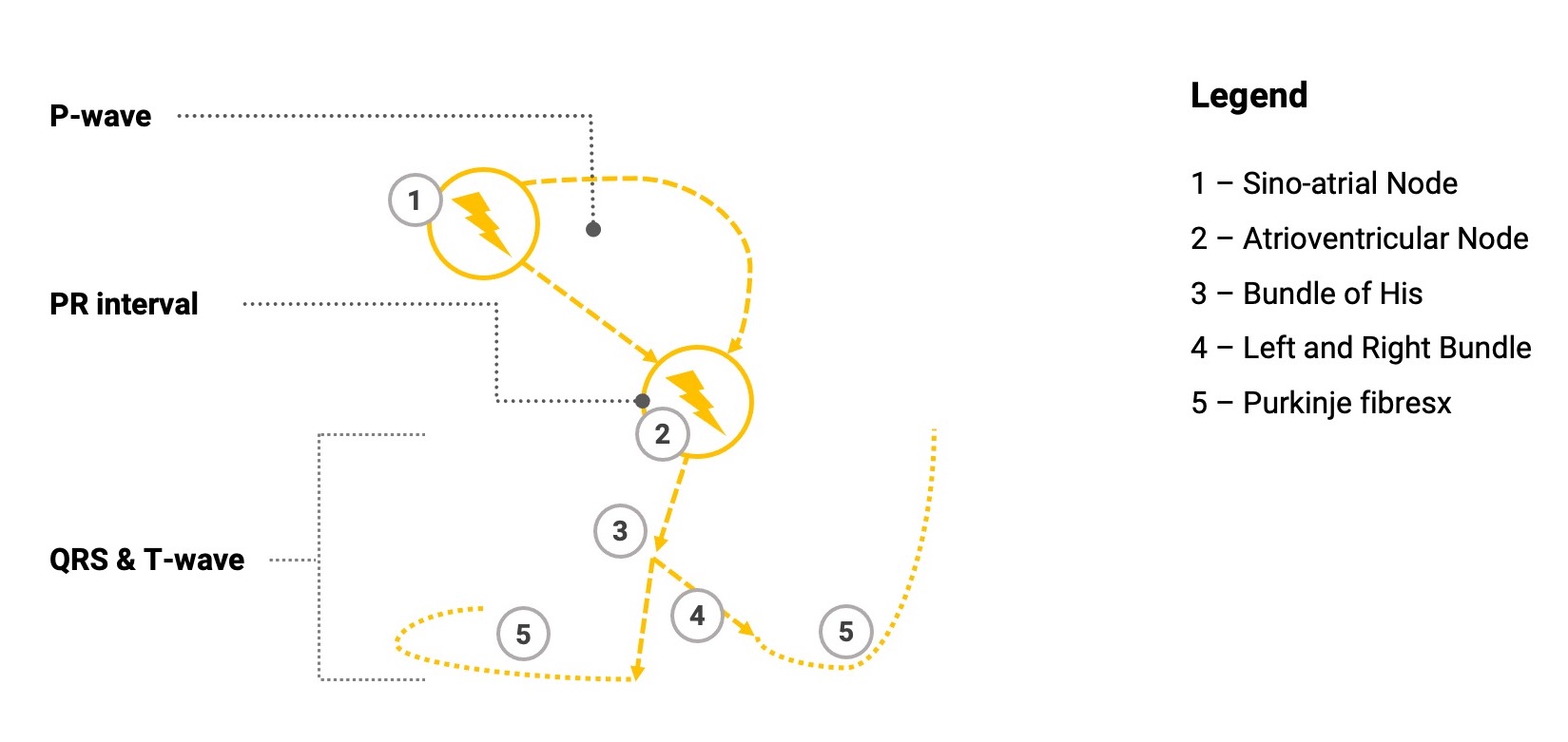
It is controlled by a conduction system which is initiated by the sino-atrial node (SAN) located in the right atrium which initiates the depolarisation and subsequent contraction of the myocytes. This is denoted by the p-wave on the ECG and represents atrial contraction. The wave of depolarisation moves across the atria where is will end up depolarising the atrioventricular node (AVN), denoted by the PR-interval. After a short delay (to allow complete contraction of the atria), the AVN depolarises and sends the action potential down into the Bundle of His, followed by the left and right bundle before reaching the Purkinje fibres which will depolarise the ventricles causing contraction, represented by the QRS complex. This is then followed by the T-wave which represents the repolarisation of the ventricles where the ventricles relax.
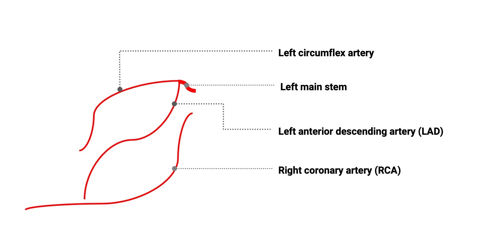
Lastly, it is supplied by a network of coronary arteries, arising from the aorta. The coronary is divided into the right and left coronary system with the right coronary artery (RCA) arising from the right cusp of the aortic valve and the left from the left coronary cusp. The RCA propagates along the heart branching out into smaller arteries. Apart from supplying the right ventricle, in the majority, the RCA provides the posterior descending artery (PDA) supplying to left ventricular posterior and inferior walls.
The left coronary system starts out with as the left main coronary artery or the left main stem. It then bifurcates into the left anterior descending (LAD) and the left circumflex artery. The LAD supplies mainly the anterior wall of the LV whilst the circumflex the lateral wall supplies the majority of the left ventricle.
In summary, the heart is a muscular pump separated into the left and right by the septa, atria and ventricle by the valves which also controls the direction of blood flow, controlled by a conduction system and supplied by the coronary arteries.